Nail Disorders
- related: Dermatology
- tags: #dermatology
The nail complex is composed of the nail plate, hyponychium, proximal and lateral nail folds, cuticle, nail bed, and matrix. The hyponychium is the skin underneath the distal free edge of the nail plate. It seals the junction between the distal nail plate and the nail bed. The nail fold protects the nail matrix and produces the cuticle. The nail bed is adherent to the bottom of the nail plate. Nail disorders can be caused by various diseases, infections, trauma, and drugs (Table 34).
Infection
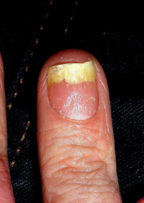
Onychomycosis is a fungal (dermatophyte) infection of the nail commonly seen in elderly persons, especially those with comorbidities such as diabetes mellitus, peripheral vascular disease, or immunosuppression. The most common sites of infection are the toenails. It can appear as distal subungual and proximal subungual onychomycosis. Distal subungual onychomycosis is the most common pattern. The distal nail plate becomes discolored (yellow, white, or brown) and thickened. Under the nail plate, there is an accumulation of subungual debris, resulting in separation of the plate from the bed (onycholysis) (Figure 141). Proximal subungual onychomycosis appears similar except that it starts at the proximal nail fold. This pattern is rare and can be associated with HIV and immunosuppression.
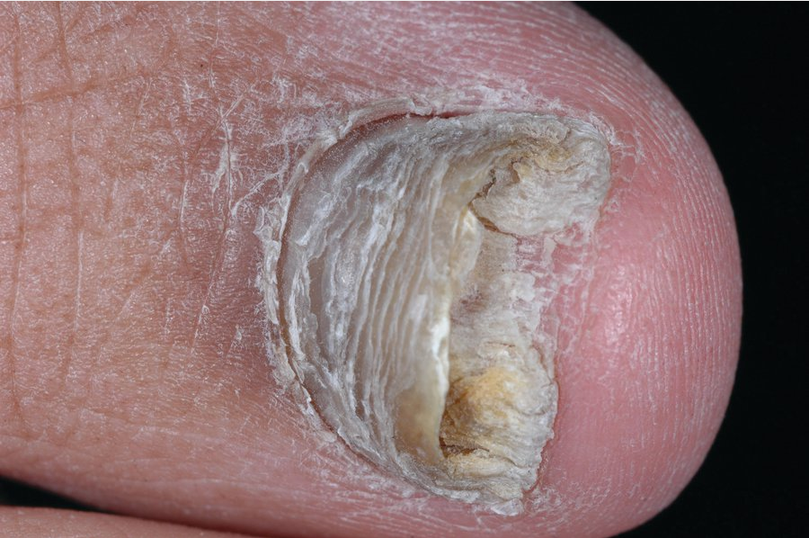
Onychomycosis is treated mostly for cosmetic reasons. Confirmation of infection with potassium hydroxide preparation, staining with periodic acid-Schiff, or fungal culture should be done before treatment. Topical antifungal agents are of limited efficacy. Oral therapy with terbinafine or itraconazole is more effective.
Paronychia
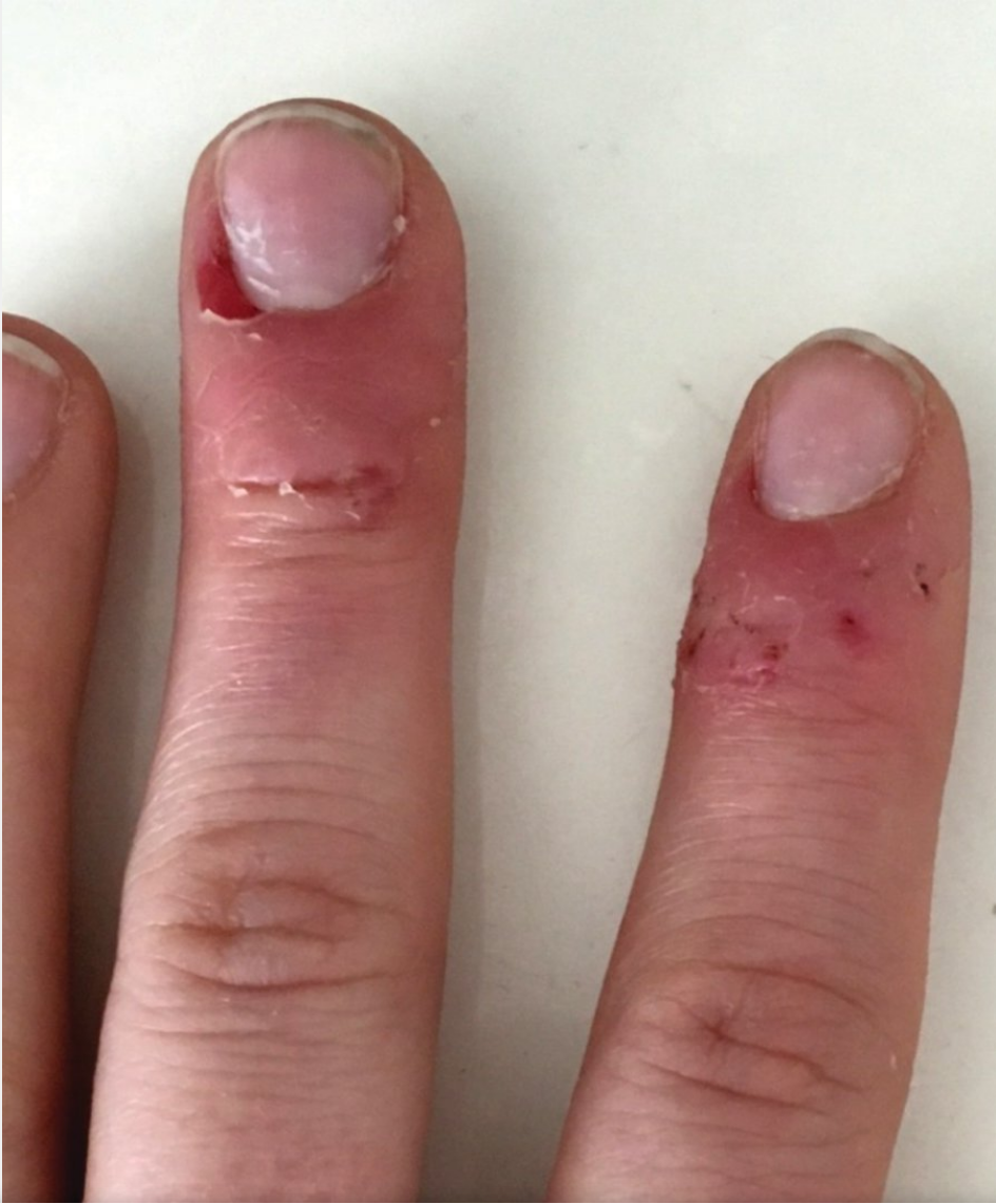
Paronychia is an infection of the nail fold. Infection is caused by trauma or chronic maceration leading to an incompetent cuticle. Acute paronychia is painful swelling of the nail fold, most commonly caused by Staphylococcus aureus. It typically affects only one nail. Chronic paronychia tends to be more insidious and involve multiple fingers. In adults, it is most often seen in those with frequent hand immersion in water or chemical contact. There is tender swelling in the nail folds with missing or dystrophic cuticles (Figure 142). Chronic paronychia can cause ridging of the nail plate. It is a multifactorial condition with several inciting factors including water, irritants, and possibly Candida species. Treatment of acute paronychia includes the use of warm compresses, incision and drainage, and in severe cases, oral antibiotics. For chronic paronychia, minimizing inciting (minimize wet work) factors is key. Treatment includes topical glucocorticoids and antifungal agents to reduce inflammation and minimizing inciting factors.
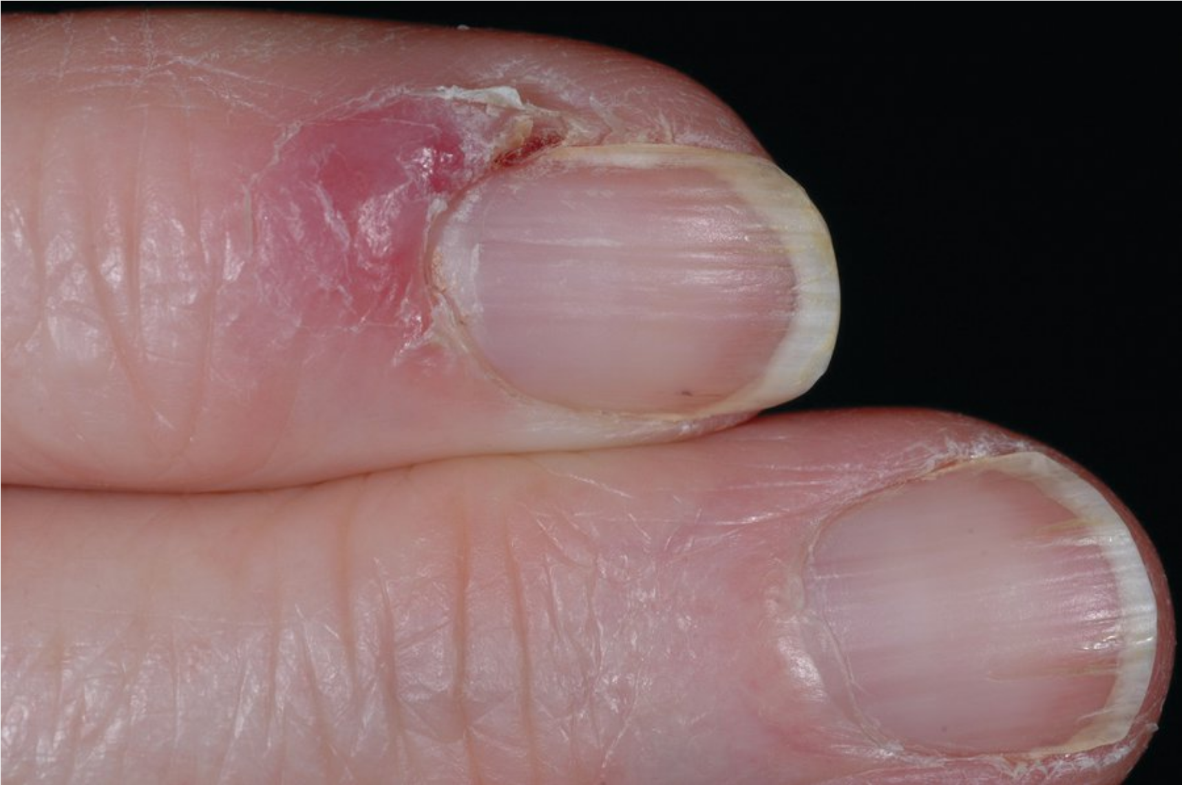 Tender, pink swelling of the proximal and lateral nail fold due to chronic paronychia. Note the dystrophic cuticle.
Inflammatory Dermatosis
Psoriasis affects fingernails more commonly than toenails. If the nail matrix is affected, tiny multiple pits will be seen on the nail plate (Figure 143). In cases of nail bed involvement, yellow-brown discoloration (oil stains) can occur. Other findings include nail plate thickening, separation of the nail plate from the nail bed, distal nail plate crumbling, and splinter hemorrhages (Figure 144). Patients with psoriatic nail involvement are often affected by psoriatic arthritis.
Lichen planus, in about 10% of cases, can affect the nails. It causes nail plate dystrophy including thinning, red-streaking, and pterygium unguis formation, which is the scarring of the proximal nail fold and matrix (Figure 145).
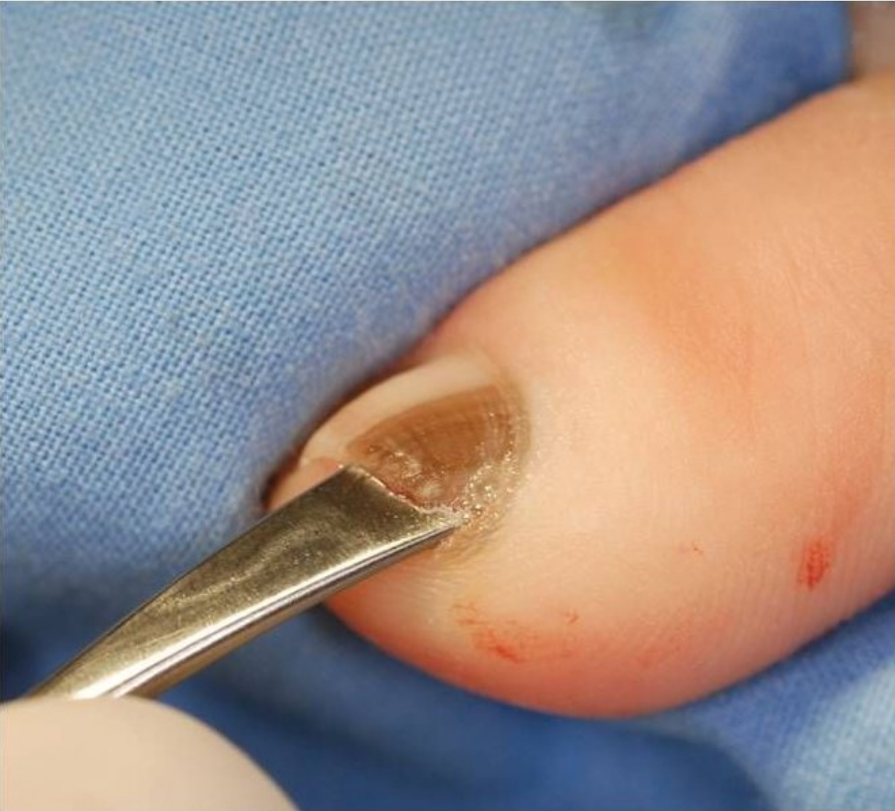
Ingrown Toenail
An ingrown toenail is the result of the nail plate growing into the lateral nail fold causing an inflammatory response. Ingrown toenails of the great toe are most common, presenting as painful swelling of the lateral nail fold (Figure 146). There may be some weeping granulation tissue.
Conservative treatment for mild to moderate ingrown nails involve wider shoes, trimming the nail plate straight across, antiseptic application, and inserting a cotton pledget under the nail edge. For severe cases, partial or complete nail avulsion with matricectomy may be necessary.
Melanonychia
Melanonychia is a brown banded pigmentation of the nail plate that can be caused by increased melanin production of the melanocytes in the nail matrix, benign hyperplasia, and melanoma (see Common Neoplasms). Longitudinal melanonychia is the transverse brown-black band on the nail plate (Figure 147), most commonly found on dark-skinned patients. Multiple nails are usually involved. Longitudinal, irregular melanonychia on a single nail can be a sign of melanoma.
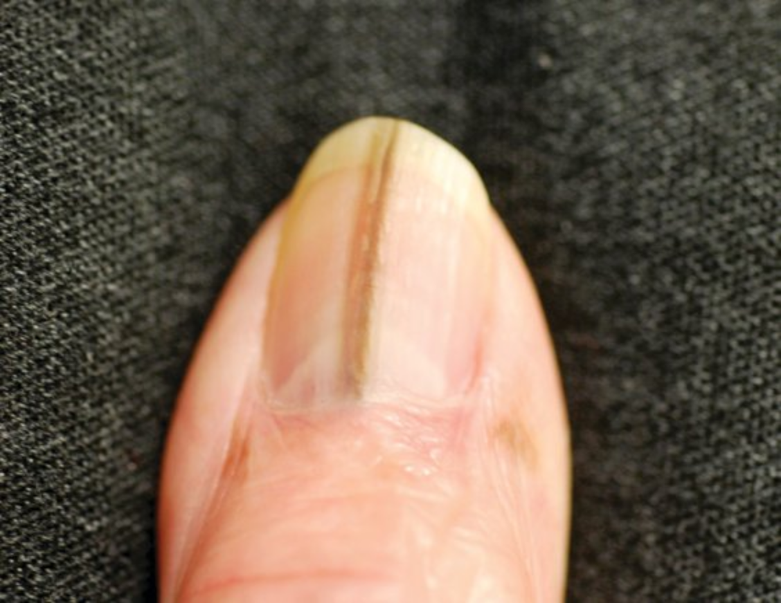
 Longitudinal brown pigmentation with transverse streaks on the nail plate characteristic of melanonychia.
Squamous Cell Carcinoma
Squamous cell carcinoma of the nail unit often presents with pain, swelling, and erythema. It commonly arises in the nail fold and appears as a hyperkeratotic papule and plaque similar to warts (Figure 148). If treatment of a wart does not progress as expected, a biopsy is warranted to rule out squamous cell carcinoma, which is strongly associated with human papillomavirus infection.
Nail pitting
This patient has nail pitting (small depressions on nail plate) and distal onycholysis (nail separation from nail bed), which are typical manifestations of psoriasis. Psoriasis is a common inflammatory skin disorder characterized by erythematous, well-defined plaques covered by thick silvery scales. The lesions are typically seen over the scalp, knees, elbows, back and gluteal cleft, and nail plates, and may be associated with mild itching. Psoriatic arthritis is a common extradermal manifestation, and the risk is increased in those with nail involvement.
The diagnosis of psoriasis is typically based on clinical findings. For this patient, a careful skin examination may identify classic plaques and clarify the diagnosis. Skin lesions are usually treated with high-potency topical corticosteroids. Mild nail psoriasis involving 1 or 2 digits can be treated with topical corticosteroids and/or vitamin D analogs (eg, calcipotriol). Widespread nail involvement, as in this patient, generally warrants systemic therapy (eg, tumor necrosis factor-alpha inhibitors, methotrexate).